Our Histology and imaging equipment
Histology/imaging equipment
Equipments for inclusion
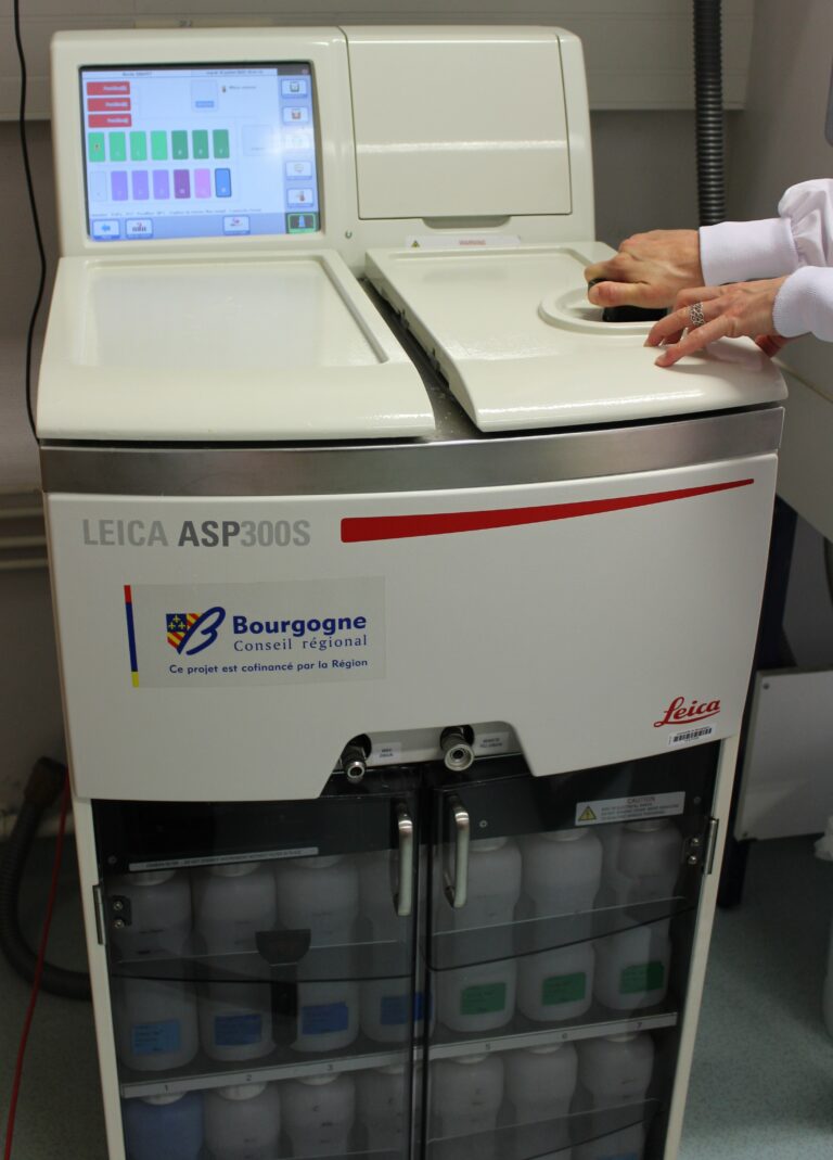
Automatic deshydration Leica ASP300
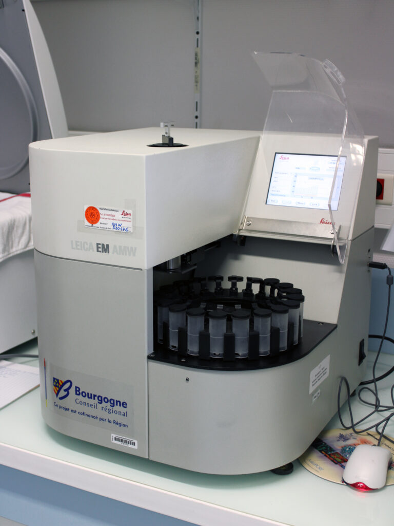
Automatic inclusion Leica EM AMW

Inclusion table Leica EG1160
Cutting equipments
Microtomes
Microtomes allow cuts from 3 µm to 40µm on kerosene blocks
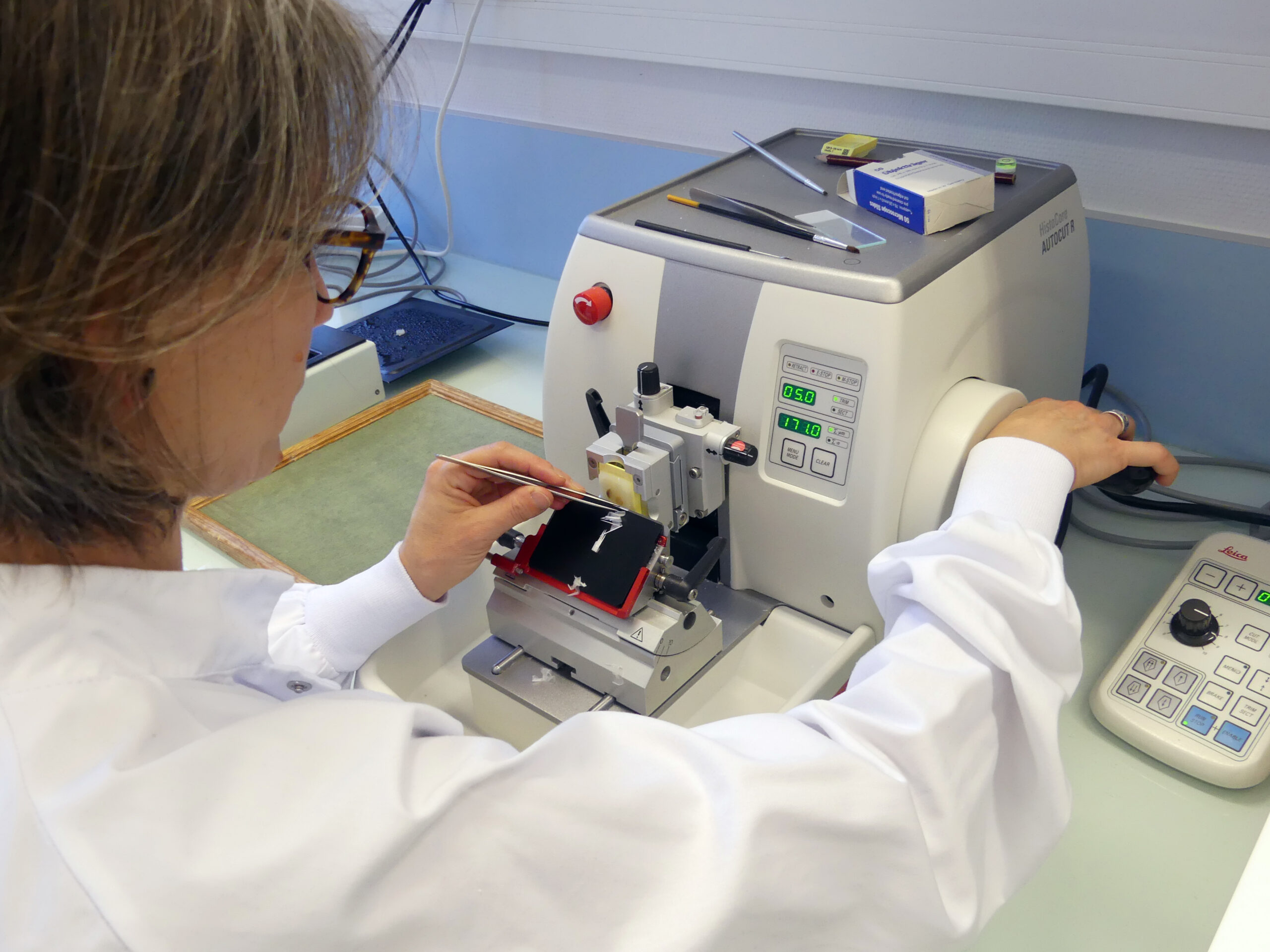
Leica Histocore AutoCut

Leica RM2145
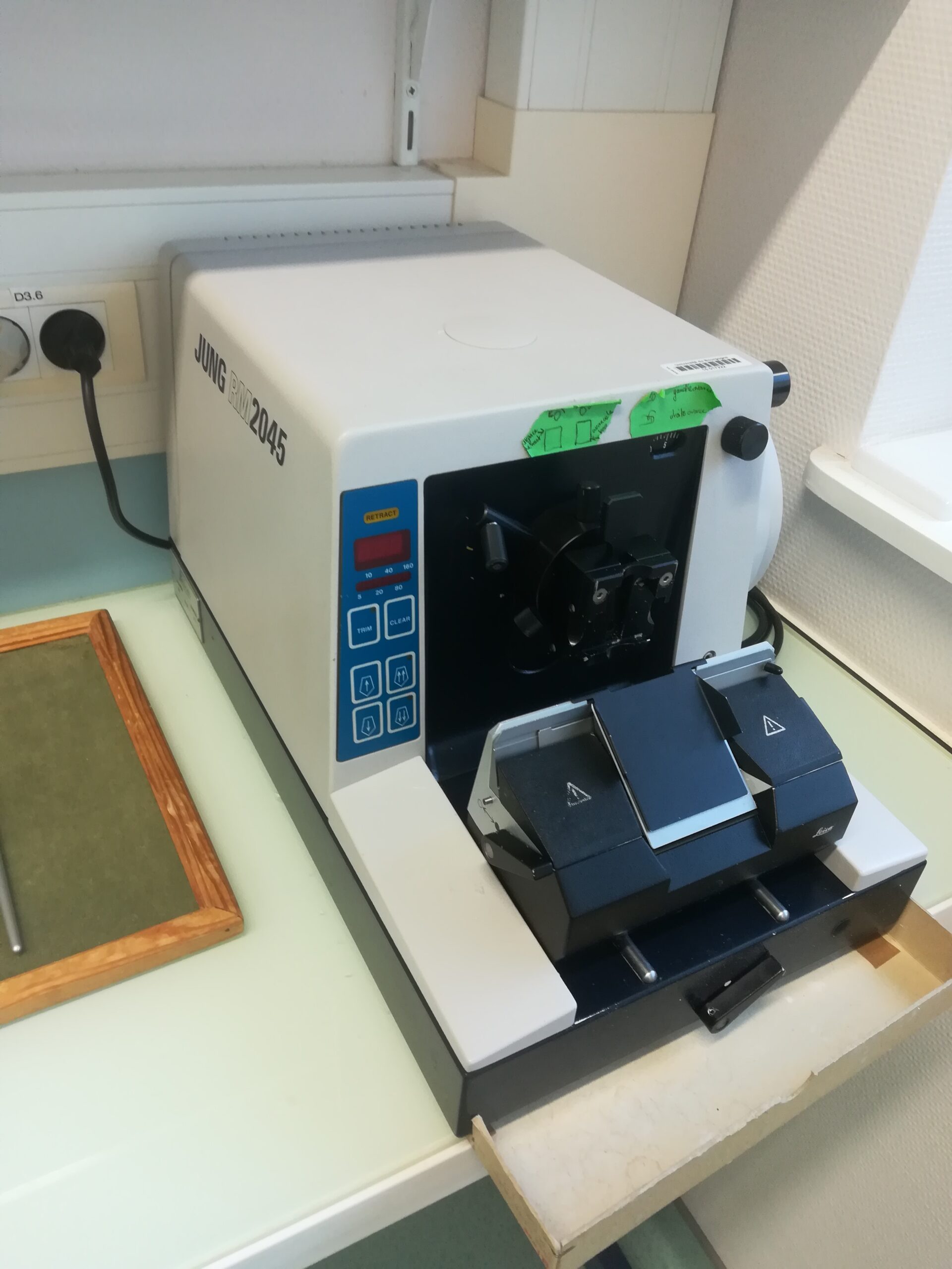
Jung RM2045
Cryostat
Cryostat allow sections from 3 µm to 40µm on frozen blocks
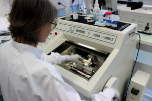
Leica CM3050S
Ultramicrotomes
Ultramicrotomes can produce semi-fine sections from 0.2 µm to 1 µm and ultra-fine sections from 60 nm to 90 nm on resin blocks.

Leica UltraCut UCT
Cutting system
Cutting system for preparing the resin block prior to ultramcrotomy.
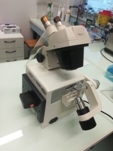
Leica EM Trim
Histological and cytological staining equipments
Autostainer
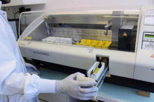
Leica Autostainer XL
Equipments for IHC ICC HIS technics
Immunostaining and in situ hybridation robot's
Leica Bond RXm
Optical microscopy equipments
Our department is equipped with a fleet of optical microscopes enabling you to acquire images at different scales and illuminations, depending on the type of support for your samples.
Microscope | Magnification | Type of support | Illumination |
Axio Zoom | x 0,70 à x 26 | Slides, plates, dishes…. | White light Fluorescence |
Axio Scope | x 2,5 à x 100 | Slides | White light Polarized light |
Axio Imager | x 5 à x 100 | Slides | Fluorescence |
Cell Observer | x 2,5 à x 40 | Slides, plates, dishes…. | White light Phase contrast Fluorescence |
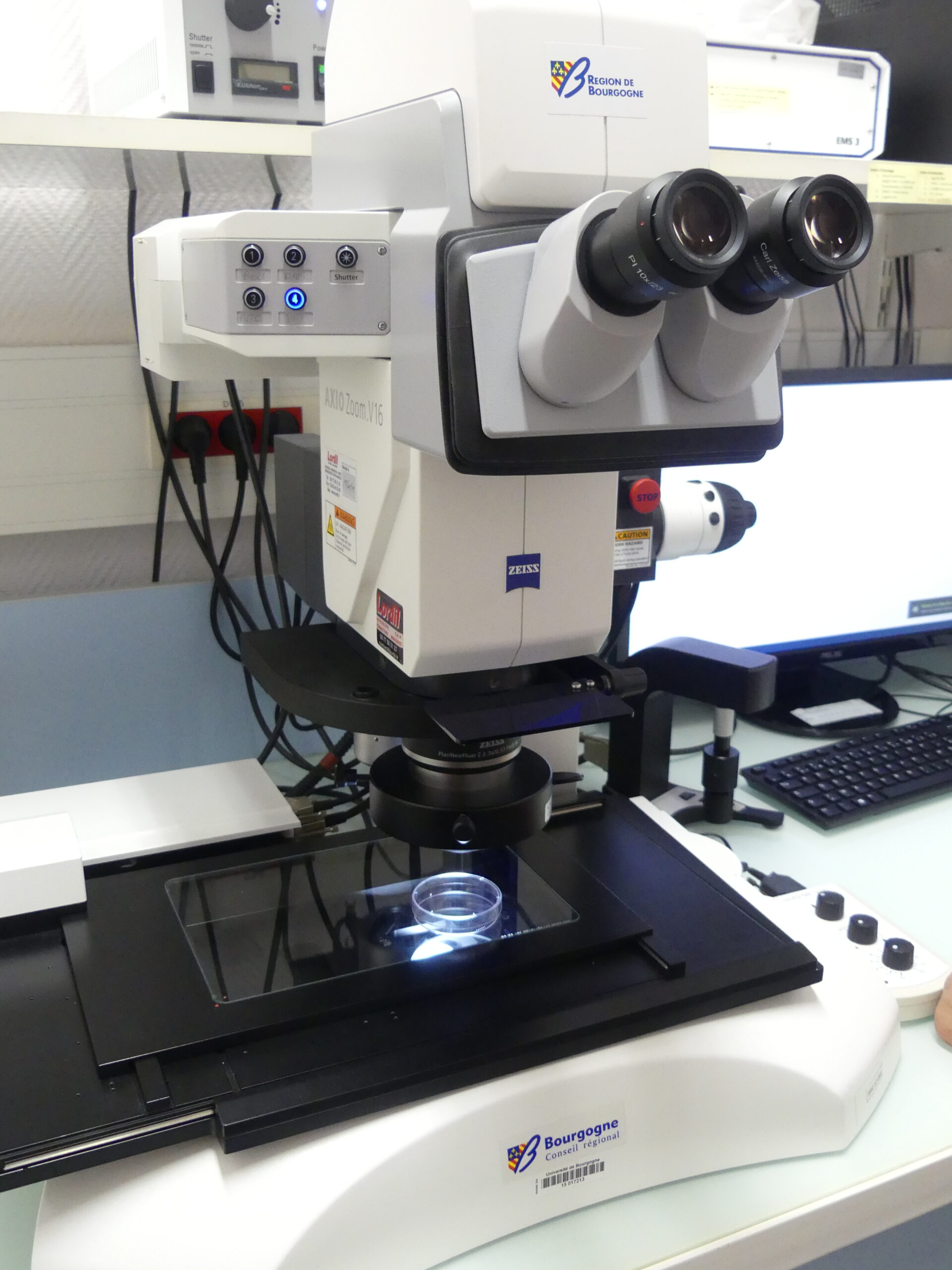
Macroscope AxioZoom (Zeiss)
Magnification : from x0.70 to x26
Illumination : Transmitted white light
Reflected white light
Fluorescence
Filters : DAPI – FITC – 63 DsRed
Applications : Mosaic, Z-stack, Time-Lapse

Microscope droit AxioScope (Zeiss)
Magnification : x2.5 – x10 – x20 – x40 – x100 oil
Illumination : White light
Polarized light
Applications : Mosaic, Z-stack
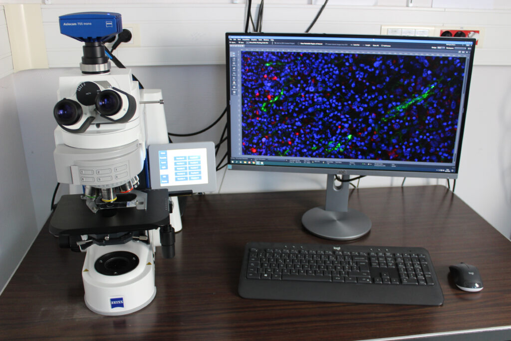
Straight Microscope - AxioImager (Zeiss)
Magnification : x2,5 – x10 – x20 – x40 – x63 oil – x100 oil
Illumination : Fluorescence
Filters: Dapi – FITC – 43 DsRed – BS640 – AF680 – BS757
Applications : Mosaic, Z-stack, Time-Lapse
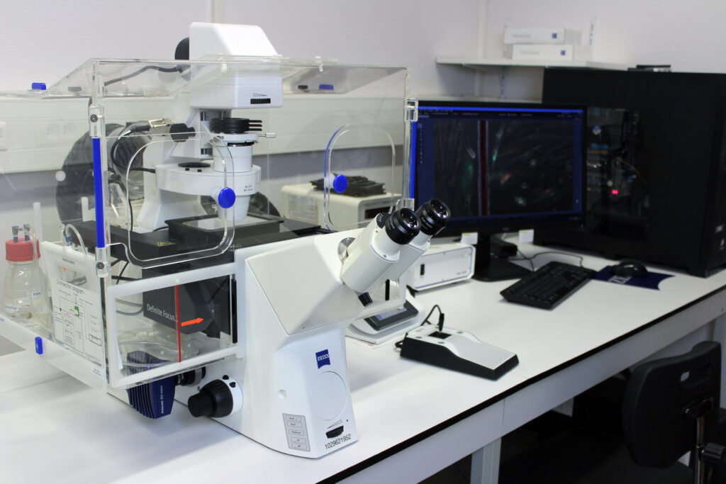
Inverted Microscope - Cell Observer (Zeiss)
The station is equipped with an incubator that maintains a controlled atmosphere in terms of CO2 and temperature, enabling live cells to be observed in real time.
Magnification : x2,5 – x10 phase – x20 phase – x40 phase
Illumination : Transmitted white light
Phase contrast white light
White light (DIC)
Fluorescence
Filtres : Dapi – FITC – 43 DsRed – 63 DsRed
Applications : long term time lapse, Mosaic, Z-stack
Detail of filters available for each microscope offering fluorescence.
|
Microscope |
Filter |
Excitation |
Emission |
|
Axio Zoom |
Dapi |
335-383 |
420-470 |
|
FITC |
450-490 |
500-550 |
|
|
63 DsRed |
559-585 |
598-660 |
|
|
Axio Imager |
Dapi |
335-383 |
420-470 |
|
FITC |
455-495 |
505-555 |
|
|
43 DsRed |
538-562 |
570-640 |
|
|
BS640 |
600-635 |
650-680 |
|
|
AF680 |
643-688 |
700-750 |
|
|
BS757 |
690-750 |
760-840 |
|
|
Cell Observer |
Dapi |
335-383 |
420-470 |
|
FITC |
450-490 |
500-550 |
|
|
43 DsRed |
532-558 |
570-640 |
|
|
63 DsRed |
559-585 |
598-660 |


